Early Cancer Detection in MRI Dataset Using a Deep Learning Mode
Cancer remains one of the leading causes of mortality worldwide, and early detection is critical to improve patient outcomes. Traditional diagnosis often relies on manual interpretation of MRI scans, which can be time-consuming and subject to human error. This project introduces an AI-driven solution that leverages deep learning algorithms, specifically convolutional neural networks (CNNs), to analyze MRI images automatically. The objective is to enhance accuracy by reducing false negatives and false positives, and to save valuable time for radiologists and clinicians, allowing earlier intervention and treatment.
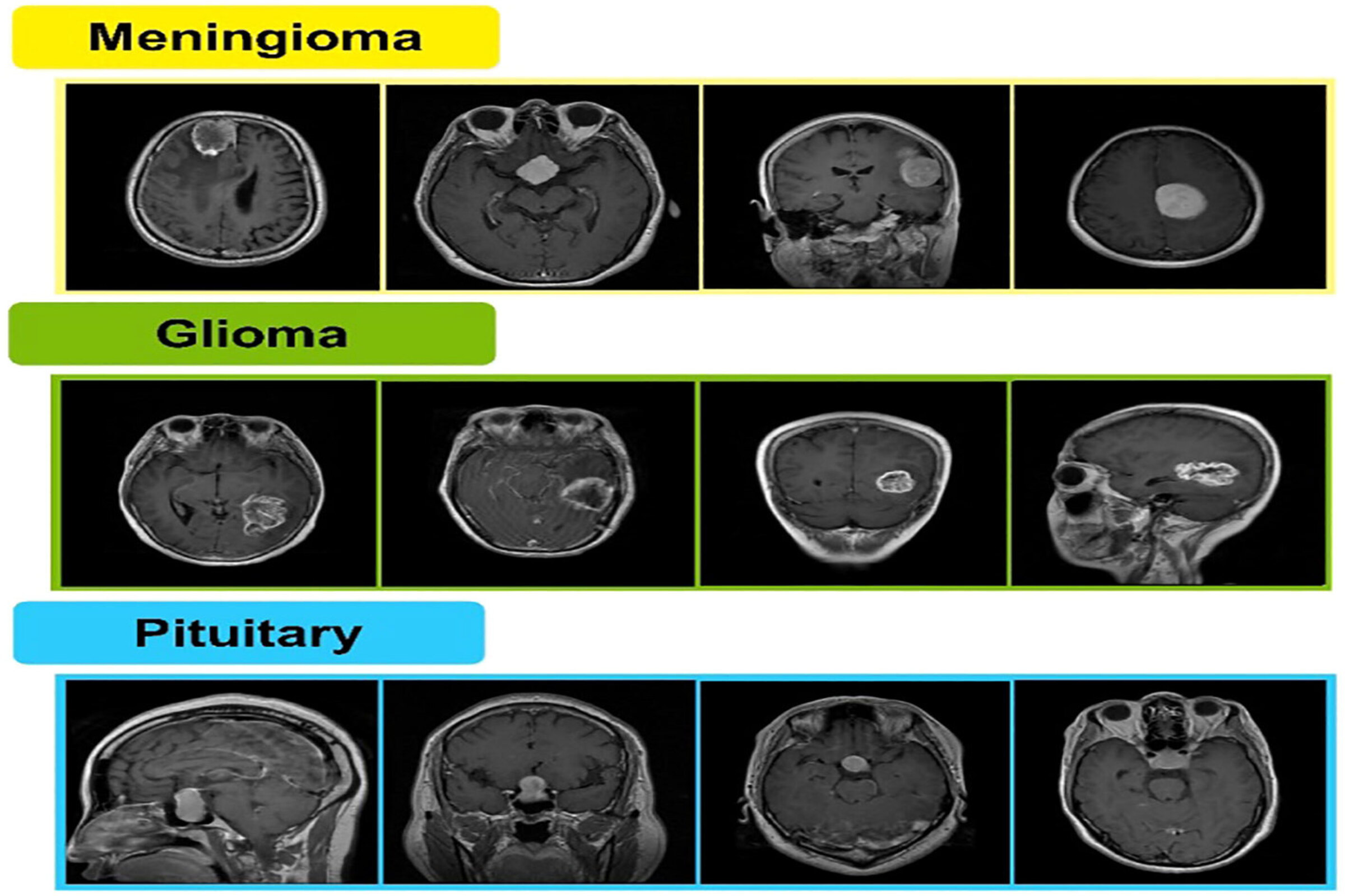
“This project is not limited to a single type of cancer. The approach can be adapted to other forms of cancer such as brain tumors, breast cancer, or prostate cancer by retraining the model with appropriate datasets. In the long term, the system can be integrated into clinical workflows to assist radiologists, reduce diagnostic delays, and standardize results across hospitals.”

The system was trained and validated on publicly available MRI datasets containing images of both normal tissues and cancerous tissues. The workflow of the project began with careful data preparation and cleaning, including the removal of low-quality or corrupted images, standardization of image formats and resolutions, and the application of augmentation techniques such as rotation, flipping, and noise injection to increase dataset diversity. Several deep learning architectures, including ResNet, VGG, and EfficientNet, were tested, and a custom CNN architecture was designed to balance computational efficiency with diagnostic accuracy. The final model achieved high sensitivity, ensuring that very few cancer cases are missed, and high specificity, reducing the risk of false alarms. These results demonstrate the reliability of the system as a supportive tool for radiologists. In addition, visualization methods such as heatmaps (Grad-CAM) were implemented to highlight the regions of the MRI that the model focuses on, improving trust and transparency for clinical adoption.